What We Do
Soft tissue surgery can include procedures to correct malformations, abnormalities in blood supply, wound management, obstructions and congenital problems, to name a few.
Surgery on the soft tissues of the body can be used to address and treat a wide range of medical conditions. Soft tissue surgery can include procedures to correct malformations, abnormalities in blood supply, wound management, obstructions and congenital problems, to name a few. Surgical oncology is often used to manage a wide variety of different cancer types and includes removal of cancers as well as reconstruction of tissues affected by either surgery or radiation treatment. Some of the more common advanced procedures involve surgery on the:
- Biliary system – gall bladder and common bile duct
- Urinary system – urethrotomy, perineal urethrostomy, cystotomy, cystopexy, kidney transplant
- Gastrointestinal system – enterotomies, end-to-end intestinal anastomosis, gastropexy , gastrotomy, splenectomy, pancreatic biopsy/pancreatectomy, liver biopsy, portosystemic shunt correction, liver mass removal
- Endocrine system – thyroidectomy, parathyroidectomy, adrenalectomy
- Respiratory system – rhinoplasty, elongated soft palate resection, laryngeal sacculectomy, lung lobectomy, tracheostomy, arytenoid lateralization for laryngeal paralsysis
- Cardiovascular system – pericardectomy, portosystemic shunt correction, vascular ring anamolies, patent ductus arteriorus, arterio-venous fistula
BAVS has available the following advanced hemostatic instrumentation:
- Ligasure
- TA stapling
- Electrocautery
Neurosurgery is performed to treat structural disorders of the brain, spinal cord or peripheral nerves.
Neurosurgery is performed to treat structural disorders of the brain, spinal cord or peripheral nerves.
Conditions of the nervous system will also have an impact on patient mobility and can be difficult to distinguish from other soft tissue or orthopedic problems. Prior to surgery, careful patient evaluation and proper diagnostics should be performed to make certain that appropriate treatment is being performed.
Diagnostics commonly necessary to correctly identify neurological problems involve advanced imaging techniques such as Myelography, Magnetic Resonance Imaging (MRI) and Computed Tomography (CT).
Neurological conditions that may require surgical treatment include: intervertebral disc herniations (IVDD), spinal fractures, “Wobblers” (caudal cervical spondylomyelopathy), lumbosacral instability/stenosis and some cancers.
Bay Area Veterinary Surgery offers a wide variety of advanced surgical procedures to manage various orthopedic conditions in canine and feline patients.
Cranial Cruciate Ligament Injury, Fractures, Hip Dysplasia, Patellar Luxation
Bay Area Veterinary Surgery offers a wide variety of advanced surgical procedures to manage various orthopedic conditions in canine and feline patients. Orthopedic procedures are designed to treat problems with structures of the muscles, tendons, bones, ligaments and joints that will lead to improved function, reduced long-term arthritis and less patient discomfort.
Cranial Cruciate Ligament Injury
One of the most common causes of lameness in the back leg of a dog is disruption of the cranial cruciate ligament (CrCL). This structure is similar to the anterior cruciate ligament (ACL) of a human and is an important stabilizer of the knee joint when an animal is standing and walking. With loss of the ligament the knee becomes unstable, leading to patient discomfort, arthritis and potential injury to an additional knee structure called a meniscus.Surgical procedures to manage loss of the CrCL are designed to improve stability to the knee joint with the intent of reducing patient discomfort, improving use of the limb, and reducing arthritis progression. Numerous procedures have been developed and should be considered for each patient on a case-by-case basis. Bay Area Veterinary Surgery offers surgical procedures such as the Tibial Plateau Leveling Osteotomy (TPLO), Tibial Tuberosity Advancement (TTA), Tightrope extra-capsular procedure, as well as others.
Fractures
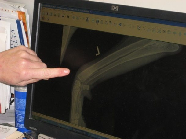
When bones break, the pieces of the bone move out of position making muscles ineffective and preventing normal movement. The broken bone also causes patient discomfort. Not all fractures require surgery, but the goals of managing a broken bone are to re-align the bone pieces as close as possible to normal (“set the bone”) and then to stabilize the bone in this position.Stabilization methods can include non-surgical methods, such as casts and splints. These methods often are not as successful in maintaining the position of the bones while they heal and require very careful monitoring and replacement, as necessary. Internal methods of stabilization “splint” directly around the bone and provide a stronger means of holding bone position.Treatment of fractures should be decided on a case-by-case basis, after careful patient evaluation, taking into consideration what will be the most effective means of returning your pet to a happy, healthy lifestyle. Surgical procedures available for fracture management include: external skeletal fixators (ESF), internal plate and screw fixation, pins, wires and interlocking nails.Many fractures in small animals are the result of blunt force trauma (hit by car) and in addition to the broken bone there can be injury to the heart, lungs, abdominal organs and soft tissues.
Hip Dysplasia
Hip dysplasia is a complex disorder with a broad range of presentations, from a patient who has difficulty walking at a very young age, to a patient who may only show problems when it is very old and suffering from the resulting arthritis. Diagnosing hip dysplasia can be difficult, and a variety of physical exam and imaging (radiographic/x-ray) techniques may be necessary to confirm a diagnosis before treatments are considered. Depending on the severity and onset of problems, a variety of medical and surgical options can be applied. BAVS offers surgical options such as Juvenile Pubic Symphsiodesis (JPS), Triple Pelvic Osteotomy (TPO), Femoral Head Ostectomy (FHO) and Total Hip Replacement (THR) as appropriate for each individual case.
Patellar Luxation
Patellar luxation is a condition where a patient’s kneecap slips out of a normal position and creates problems. Patients with patellar luxation suffer a loss of muscle strength, discomfort and ultimately can form arthritis in the knee joint. Patellar luxation is one of the least completely understood and more complex orthopedic disorders that is managed in companion animals. There are multiple predisposing factors that have to be assessed and addressed for each patient. Ultimately, surgery is aimed at restoring the normal anatomic position of the kneecap and then stabilizing the kneecap in that position. Surgical procedures can include, but are not limited to: trochleoplasties, tibial tuberosity transpositions, imbrications and desmotomies. Advanced procedures such as distal femoral osteotomies can also be employed as necessary.
Minimally invasive surgery techniques have evolved to permit visualizing and working on internal structures using magnification and smaller instruments.
Arthroscopy, Laparoscopy and Thoracoscopy
Traditional surgery involves visualizing and working on internal structures through relatively large incisions. Minimally invasive surgery techniques have evolved to permit visualizing and working on internal structures using magnification and smaller instruments. Advantages include:
- Smaller incisions
- Less trauma
- Less patient discomfort
- Improved examination of structures
- Faster recoveries
- Shorter hospitalization times
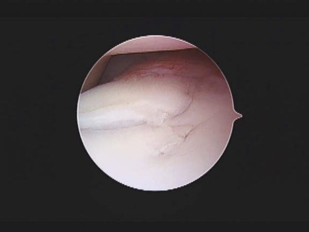
Arthroscopy (Joints)
The canine shoulder joint’s size and anatomy make it particularly suitable to arthroscopic procedures.Arthroscopy involves making a small incision around the joint (shoulder, elbow, knee, hip) and inserting a small endoscope camera (arthroscope) into the joint to examine the internal structures. Additional small incisions are made as necessary to insert instruments to allow for the surgeon to work on the joint. This approach allows for many surgical problems of joints to be diagnosed or treated with less patient discomfort and a faster recovery.
Conditions of the shoulder joint that can be diagnosed or managed with arthroscopy include: osteochondrosis desicans (OCD), bicipital tenosynovitis, joint instability as well as diagnosis of arthritis and cancers.
Conditions of the elbow that are commonly evaluated with arthroscopy include: “elbow dysplasia” from fragmented medial coronoid processes (FCP), osteochondrosis desicans (OCD), ununited anconeal process (UAP), incongruency conditions and diagnosis of arthritis and cancers.
Evaluation of the hip joint helps to identify and treat some tears in the labrum (cartilage) and assist in the overall diagnosis and management of hip dysplasia/hip arthritis.
Evaluation of the stifle is commonly performed to treat osteochondrosis desicans (OCD), to confirm a diagnosis of a partial tearing of the cranial cruciate ligament (animal version of the ACL in a human), damage to the meniscus or origin of an extensor tendon (long digital extensor). It can also be used to help diagnose arthritis and biopsy cancers.
Thoracoscopy (Chest Cavity)
Thoracoscopy allows for a surgeon to work on many lung, heart and esophageal structures through smaller incisions instead of “cracking” the chest wall. Thoracoscopy improves patient recovery and leads to less patient discomfort. Procedures can include: acquisition of biopsies, removal of portions of the pericardium (sac around the heart) to manage pericardial effusion and diagnosis/management of some causes of blood (hemothorax), air (pneumothorax) or other fluids (chylothorax) in the chest.
Laparoscopy (Abdominal Cavity)
Laparoscopic or laparoscopic-assisted procedures are successful in treating a wide variety of abdominal problems without a need for a larger abdominal incision. Procedures include: liver biopsies, biopsies of other abdominal organs (kidneys, small intestines), assistance in bladder surgery/bladder stone retrieval, prophylactic gastropexy (surgery performed as a preventative measure in the management of “bloat”), spays and retained testicle castration (cryptorchid).
Just as digital cameras have replaced film-use cameras for most of us, digital radiography or x-rays have largely replaced the use of radiographic film in imaging.
X-Rays
Just as digital cameras have replaced film-use cameras for most of us, digital radiography or x-rays have largely replaced the use of radiographic film in imaging. Since there is no film to process and store, digital images can be easily accessed almost immediately on a computer screen by multiple care providers. Digital radiography allows for greater image clarity and flexibility in sharing information with your family veterinarian and the entire animal care team.
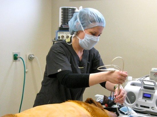
A licensed veterinary technician (LVT) has a college degree in veterinary technology and has passed a national board examination demonstrating specific knowledge and competencies in both high-quality nursing techniques and high-risk anesthesia.
Managing Your Pet's Pain
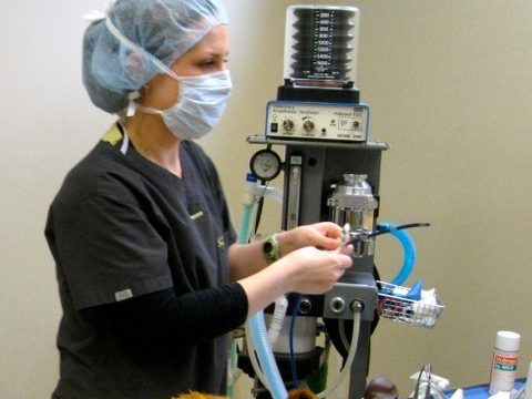
Nursing expertise is a very important part of anesthetic safety. A licensed veterinary technician (LVT) has a college degree in veterinary technology and has passed a national board examination demonstrating specific knowledge and competencies in both high-quality nursing techniques and high-risk anesthesia. These individuals are licensed by the state and must fulfill annual continuing education requirements.
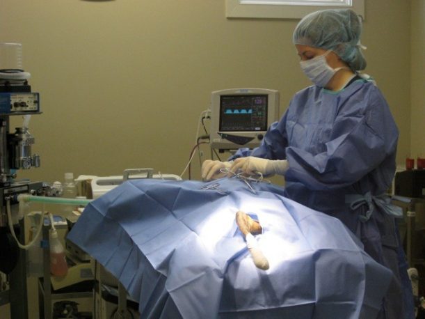
Our LVTs, under the guidance of the surgeon, oversee anesthesia for every surgery to ensure the highest quality of care for our patients. This monitoring allows our surgeons to focus more on the surgical procedure, reducing the risk of surgical and anesthetic complications. All of our surgical patients have a physical exam, pre-anesthetic blood work, and are connected to anesthetic monitors that continuously measures oxygen levels, ventilation status, heart rate and rhythm (EKG), respiratory rate, body temperature and blood pressure. In addition, every anesthetized patient receives supportive IV fluid containing pain relievers to maintain blood pressure as well as active and passive warming to maintain body temperature.All tissue and bone injury, whether it be from trauma, elective surgery or an ongoing medical condition, can cause pain and affect quality of life. The goal of many surgical procedures is to ultimately improve quality of life and alleviate pain, but it is true that while healing many patients experience discomfort.
Managing your pet’s pain is an important part of our process, and we incorporate both pharmacologic and non-pharmacologic techniques to anticipate and prevent as much discomfort as possible. Each patient receives narcotic pain relievers before anesthesia, during anesthesia and typically for the first night following anesthesia. We also employ local anesthetics when indicated.
At Bay Area Veterinary Surgery we are committed to providing exceptional care. We want to ensure that each and every patient has a good experience by treating them as if they are our own.